Computed, not scripted responses.
The physiology engine is the architectural core of the full-system mannequin. Most training simulators play back a scenario: a pre-recorded sequence of vitals with branch points a technician triggers. This engine runs a model instead. Named physiological quantities - blood volume, cardiac output, sympathetic tone, drug plasma concentration, and more - are connected by real equations and recalculated numerous times every second. No clinical outcome is ever written into the code as a rule. It has to emerge from the underlying variables, or it doesn't happen.
ODE-Based Cardiovascular Modeling
Heart rate, stroke volume, cardiac output, and systemic vascular resistance are continuously solved. Interventions shift the state - the engine propagates the consequences forward.
Mechanism-Level Pharmacology
Drug interactions emerge naturally from the model rather than being hard-coded. Every drug acts only on named physiological mechanisms - contractility, chronotropy, SVR, respiratory drive. If a trainee gives atropine for a bradycardia caused by hypoxia, the heart rate doesn't come up, because atropine and hypoxic bradycardia work through two different pathways in the model.
Respiratory and Autonomic Coupling
Respiratory mechanics, oxygenation, and CO₂ clearance are modeled alongside cardiovascular state and other parameters. Autonomic compensatory responses emerge from the coupled system.
RECOVER-Guideline CPR Validation
The primary training scenario is validated against RECOVER CPR guidelines, built into the engine directly. Compression depth, rate, and chest recoil are sensed and scored - efficacy directly influences perfusion pressure in the model.
The math under the hood.
Every parameter is calculated in real time using industry accepted mathematical expressions.
Frank–Starling Stroke Volume
Stroke volume saturates with filling pressure and falls as afterload rises. A real saturating curve recomputed every 100 ms, not a lookup table.
Baroreflex Lag
One ODE is the entire baroreflex: a first-order lag toward a sigmoid target of blood-pressure error. Why heart rate climbs as pressure falls, and why the reflex exhausts in decompensated shock.
Hill / Emax Dose–Response
Every drug's effect on every physiological hook uses this same saturating binding curve. A new drug is a new set of numbers in a catalog file. The solver never changes.
Findings you can actually feel and see.
Physical exam findings on the mannequin are driven by the physiology engine - not preset states. What the trainee detects depends on where the patient is physiologically at that moment.
Palpable Pulses
Bilateral femoral, dorsal pedal, and lingual pulse sites. Pulse character, rate, and rhythm reflect real-time cardiac output. Pulse deficits and arrhythmia-associated changes are physically detectable.
Auscultatable Heart & Lung Sounds
Embedded speakers deliver cardiac and pulmonary sounds that reflect the physiology engine state in real time. Asynchronous rhythms - like PEA vs. VF - are distinguishable by auscultation vs. pulse palpation.
Peripheral Temperature Gradient
Extremity temperature differs from core temperature as a physical finding in low-perfusion states. Sensor-confirmed readings at standard clinical assessment sites.
Ocular Responses
Pupillary light reflex and menace response are simulated. Pupil dilation state and PLR reactivity reflect the neurological status computed by the physiology engine.
Mucous Membrane Color
Dynamic MM color display reflects modeled perfusion, oxygenation, and shock state - transitioning from pink through pale, white, and cyanotic or icteric as appropriate to the scenario.
Respiratory Chest Rise
Internal bladder inflation synchronized to the modeled respiratory rate produces visible chest rise. Rate and quality change with clinical interventions and physiological deterioration.
Train with the same tools you use in practice.
No proprietary adapters. No simulation-only equipment. Standard clinical tools work on the mannequin without modification - eliminating the muscle memory mismatch between simulation and clinical practice.
Physical CPR with real-time feedback - validated against RECOVER guidelines
Compression depth, rate, and chest recoil are sensed and scored during CPR. Efficacy directly influences modeled perfusion pressure and resuscitation outcome. Instructor and trainee see performance metrics in real time during the scenario - and can review them in debrief. The physiology engine means a given compression sequence produces different outcomes depending on the patient's state at the time.
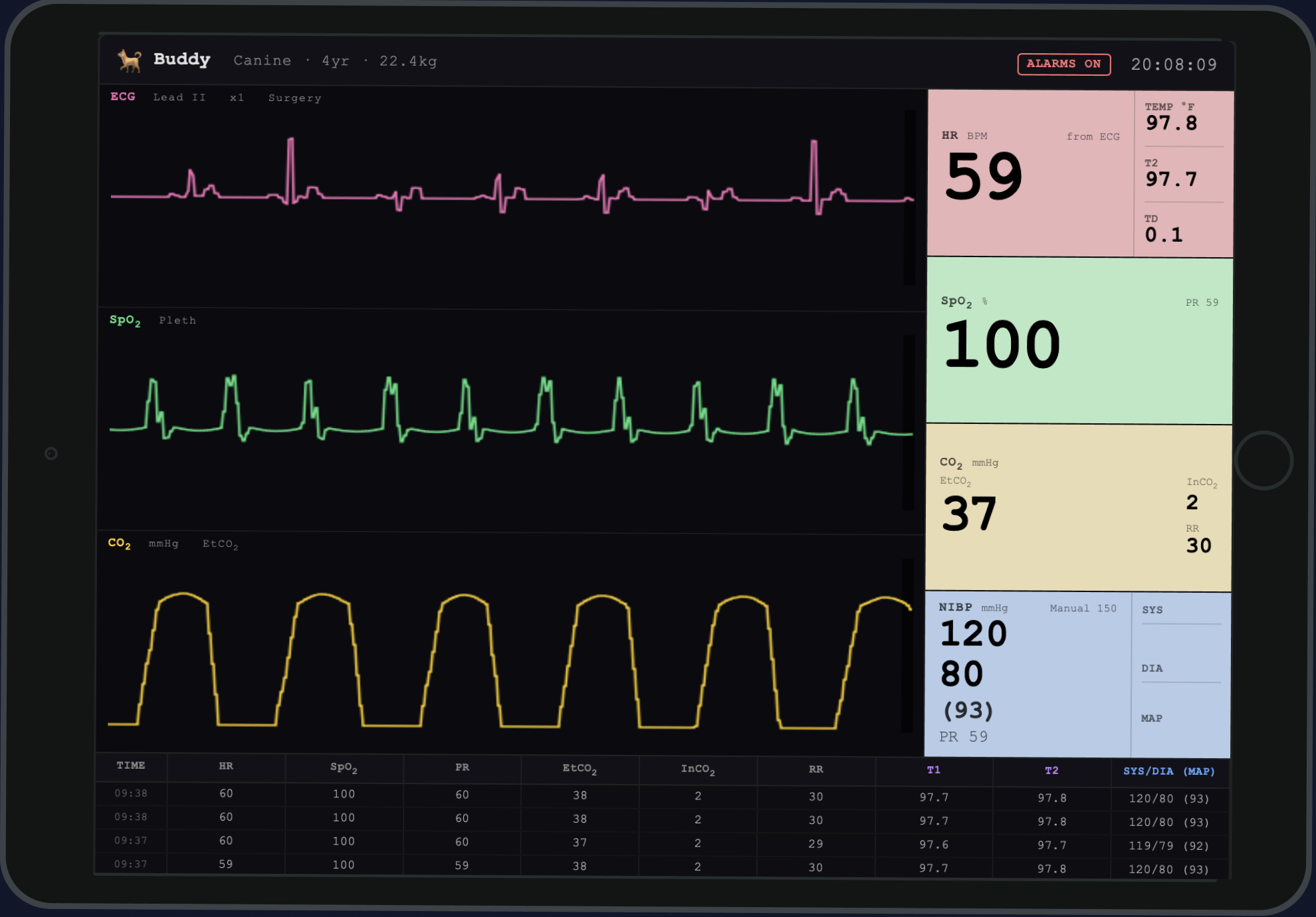
A monitor that looks familiar.
The simulated patient monitor replicates the interface and waveforms trainees encounter in clinical practice. Trainees learn to interpret a display that is similar in structure and layout to many common veterinary telemetry machines. Every value on screen is live output from the physiology engine.
- ECG Live waveform reflects engine state: NSR, brady, tachy, PEA, asystole, VF
- SpO₂ Waveform and numeric output from modeled oxygenation
- ETCO₂ Capnography waveform morphology changes with ventilation quality and cardiac output
- BP Doppler audio simulation for manual technique; numeric on monitor
- TEMP Core temperature numeric; differs from peripheral findings
Standalone skill models for high-repetition practice.
Each trainer is built with medical-grade silicone for tactile and anatomical fidelity. Compatible with standard clinical equipment - no proprietary tools required. Available as catalog models or custom builds.
NGT Placement Model
Canine head and esophageal model with realistic resistance at the nasopharynx and gastroesophageal junction. Confirms correct gastric placement and supports feeding tube and decompression tube practice. ET tube placement is planned for V2.
PICC Placement Limb Model
Peripherally inserted central catheter placement via the saphenous or cephalic approach, with depth and advancement cues confirming central line position. Pressurized vascular system produces blood flash on vessel entry. Also supports standard IV and IO placement, though PICC is the primary use case. Repeated use with replaceable vessel inserts.
Chest Wall Model
Bilateral pleural space compartments with fluid and gas character feedback. Compressible chest wall with anatomically appropriate intercostal spacing. Supports both needle and tube approaches.
Subxiphoid Model
Fluid-filled pericardial compartment with tamponade simulation. Anatomically referenced depth and resistance cues for the subxiphoid approach. Supports pericardial sac puncture and aspiration.
Abdominal Wall Model
Layered abdominal wall with fluid-filled organ analogs. Supports abdominocentesis in a four-quadrant approach and cystocentesis with bladder puncture and aspiration.
Ultrasound-Compatible Phantom
Gelatin/silicone composite construction for ultrasound signal fidelity. Hepatorenal, splenorenal, pericardial, and urinary views. Includes mock fluid collections for positive-finding training.
Built to your program's specifications.
DVMiQ Simulation accepts custom build requests from veterinary institutions, residency programs, and educators. Custom projects begin with a scoping consultation.
- Species and size variation - feline, exotic, pediatric canine
- Procedure-specific trainers outside the current catalog
- Integrated sensors and data logging for assessment programs
- Institutional branding and anatomical labeling
- Multi-unit production runs for skills lab deployment
Inquiry - Early Access & Custom
Or email us directly at support@dvm-iq.com
The physiology engine is actively undergoing automated & rigorous testing by veterinary professionals. A formal face-validity review with board-certified veterinary criticalists will be required before release. DVMiQ Simulation is not yet available for purchase - institutions and practices interested in early access or pre-release partnership are encouraged to reach out now.
Interested in early access?
DVMiQ Simulation is taking pre-release inquiries for both the hardware mannequin and DVMiQ Sim Online, from veterinary schools, residency programs, clinical skills labs, and individual clinicians.